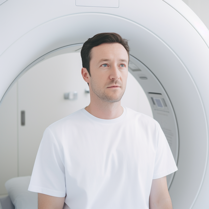
An MRI, or magnetic resonance imaging, is a medical imaging technique used to produce detailed images of body tissues and organs. It is a non-invasive procedure that uses a powerful magnetic field and radio waves to generate images of the inside of the body.
The purpose of an MRI is to diagnose a wide range of conditions, including cancers, brain tumors, spinal cord injuries, joint abnormalities, and other disorders. The machine creates images by aligning the magnetic moments of hydrogen atoms in the body using a strong magnetic field. Radio waves are then used to disturb the alignment of these atoms, and the energy released as they realign is detected and used to construct detailed cross-sectional images of the body.
An MRI can produce various types of images, including T1-weighted, T2-weighted, and fluid-attenuated inversion recovery (FLAIR) images. These images can provide valuable information about the structure and function of tissues and organs, allowing healthcare professionals to make accurate diagnoses and develop appropriate treatment plans.
In summary, an MRI is a powerful medical imaging tool that uses a combination of magnetic fields and radio waves to create detailed images of body tissues and organs, making it an invaluable tool for diagnosing a wide range of medical conditions.
When preparing for an MRI, there are several important considerations to keep in mind to ensure the procedure goes as smoothly as possible. From understanding the purpose of the MRI to pre-appointment preparations and what to expect during the actual scan, being well-informed and prepared beforehand can help alleviate any anxiety or concerns. It's important to follow any specific instructions given by the imaging facility and communicate openly with your healthcare provider about any potential concerns or pre-existing conditions that may affect the imaging process. By taking the time to properly prepare for an MRI, individuals can help ensure a successful and comfortable experience.
Before undergoing an MRI scan, it is crucial to remove all metal objects to ensure the safety and accuracy of the procedure. This includes jewelry, hairpins, zippers, body piercings, watches, hearing aids, pocketknives, and eyeglasses. Any metal item can interfere with the magnetic field of the MRI scanner, causing distortion in the images and potentially posing a safety risk to the patient.
If you have any metal inside your body, such as implants or pacemakers, it is essential to inform your doctor or the technologist before the MRI scan. They will assess whether the test can be conducted safely or if alternative imaging methods should be considered.
Failure to remove metal objects or inform the medical staff about internal metal may result in serious consequences, including burns, tissue damage, or movement of implanted devices. It is important to adhere to these guidelines to ensure the success and safety of the MRI scan.
In conclusion, prior to undergoing an MRI scan, it is crucial to remove all metal objects and inform the medical staff about any metal inside your body to ensure the safety and accuracy of the procedure.
Before undergoing an MRI, it is essential to inform the technologist about any medical conditions, allergies, or health concerns that could interfere with or pose a risk during the procedure. This includes but is not limited to, implanted devices such as pacemakers, defibrillators, or cochlear implants, as the strong magnetic fields used in MRI can affect these devices. Additionally, the presence of metallic objects in the body, such as shrapnel or surgical implants, can also pose a risk and should be disclosed to the technologist.
Other medical conditions such as claustrophobia, anxiety disorders, or severe obesity may require special accommodations or considerations during the MRI. Allergies to medications, contrast dyes, or other substances that may be used during the MRI should be communicated to the technologist to prevent any adverse reactions.
By providing detailed information about medical conditions, allergies, implanted devices, and metallic objects, the technologist can take the necessary precautions to ensure the safety and well-being of the patient during the MRI procedure.
To change into a hospital gown, first remove all clothing and undergarments. Ensure that you are completely undressed before putting on the hospital gown. Then, put on the hospital gown with the opening in the back. Make sure the gown is positioned properly and the opening is at the back.
Next, tie the gown securely to ensure it stays in place. The ties should be fastened tightly enough to prevent the gown from coming loose, but not so tight that it causes discomfort. Check that the gown is comfortable to wear and does not restrict movement or cause any discomfort to the individual.
It is important to follow these steps to ensure the hospital gown is worn correctly and comfortably. By removing all clothing and undergarments and putting on the gown properly with the opening in the back, the individual can feel secure and comfortable during their time in the hospital.
The interaction between the Earth's magnetic field and radio waves is a complex and fascinating topic. Understanding how these two phenomena are intertwined is crucial for a variety of technological applications and scientific research endeavors. In this exploration, we will delve into the nature of the Earth's magnetic field, its influence on the behavior of radio waves, and how scientists and engineers utilize this knowledge in various fields such as communication, navigation, and space exploration. Additionally, we will discuss the impact of the Earth's magnetic field on radio wave propagation, including the concept of magnetic storms and the potential effects on communication and navigation systems. By examining the relationship between the magnetic field and radio waves, we can gain a deeper appreciation for the interconnectedness of these fundamental aspects of the natural world and their significance in modern society.
MRI machines work by utilizing a powerful magnetic field, radio waves, and computer processing to create detailed images of the body. The patient lies on a table that slides into a cylindrical machine containing a strong magnet. The magnetic field aligns the protons in the body's soft tissue. When radio waves are directed into the body, the protons become excited and emit their own, which are then detected by the MRI machine. These emitted signals are organized into detailed images by the computer.
The essential components of an MRI machine include the table, cylinder, magnet, and radiofrequency coils. The table allows for precise positioning of the patient within the machine, while the cylinder houses the powerful magnet. Radiofrequency coils are used to send and receive the radio waves necessary for the imaging process.
In summary, MRI machines work by using a strong magnetic field to align the protons in the body's soft tissue, then stimulating these protons with radio waves and detecting their emitted signals to create detailed images through computer processing.
The magnetic field is a force field that surrounds a magnet and is created by the movement of electric charges. It has the ability to attract certain materials, such as iron and steel, and repel others. This is due to the alignment of the magnetic field lines in the presence of another magnetic field. The Earth's magnetic field plays a crucial role in protecting the planet from harmful solar winds and cosmic radiation, creating a magnetosphere that shields the atmosphere.
In everyday life, the magnetic field has various practical applications. For example, it is used in compasses for navigation, in MRI machines for medical imaging, and in speakers and headphones for sound reproduction. It is also used in magnetic levitation trains and in the generation of electricity in power plants. These examples demonstrate the wide-ranging impact of the magnetic field in modern technology and daily life.
Radio waves are used in medical imaging, particularly in MRI scans, to create detailed images of the body's internal structures. MRI machines rely on the behavior of hydrogen atoms when exposed to radio waves. When the body is placed in a strong magnetic field, these atoms align with the field and can be further manipulated by radio waves. As the atoms return to their natural alignment, they emit radio signals that are used to create detailed cross-sectional images of the body's tissues and organs.
In the formation of synthetic aperture radar (SAR) images, radio waves play a crucial role in remote sensing and geological mapping. SAR systems transmit radio waves towards the Earth's surface and then capture the waves that are reflected back. By measuring the time it takes for the waves to return and how they have been altered, SAR can create high-resolution images of the Earth's surface, which are valuable for various applications including monitoring environmental changes, disaster management, and resource exploration.
In both medical imaging and remote sensing, the use of radio waves has revolutionized the ability to create detailed and accurate images of the internal structures and surfaces, allowing for advancements in healthcare, environmental monitoring, and geological mapping.
An MRI (Magnetic Resonance Imaging) machine is a powerful medical tool that uses a magnetic field and radio waves to create detailed images of the inside of the body. The process involves laying inside a large cylindrical tube, which can be intimidating for some patients. Understanding how the MRI machine works, what to expect during the procedure, and how to prepare for it can help alleviate anxiety and ensure a smooth experience for patients. In this article, we will explore what it's like inside the MRI machine, including the technology behind it, the positioning and comfort of the patient, and the importance of remaining still during the imaging process. We'll also discuss how to mentally and physically prepare for an MRI scan, as well as provide tips for staying calm and comfortable while inside the machine.
To enter the machine, there are several necessary steps that must be followed to ensure safety and proper operation. Before entering the machine, it is important to wear the recommended attire, including a hard hat, safety goggles, and appropriate clothing such as long pants and closed-toe shoes for protection.
Once properly dressed, the first step is to thoroughly inspect the machine for any potential hazards or malfunctions. It is crucial to ensure that the machine is turned off and locked out to prevent any accidents while entering.
Next, carefully approach the machine and use the designated entry point, following any specific instructions for accessing the interior. Take note of any warning signs or labels and proceed with caution.
Before entering the machine, it is important to communicate with any other workers in the area to ensure that everyone is aware of the machine entry and to prevent any unexpected movements or operations.
Once inside the machine, it is important to be aware of any potential hazards and to follow all safety precautions while performing the necessary tasks.
Overall, the process of entering the machine requires careful attention to safety precautions, proper attire, and thorough communication with other workers to ensure a safe and successful entry.
An MRI scan is an important medical imaging tool that allows doctors to obtain detailed and accurate images of the body's internal structures. Staying still during the MRI scan is crucial to ensure that the images obtained are clear and accurate. Even the slightest movement can cause blurriness or distortion in the images, compromising the quality of the results.
The potential consequences of moving during the MRI scan can have a significant impact on the diagnostic accuracy. It can lead to the need for repeat scans, which can be time-consuming and costly. More importantly, it may result in missing important details or abnormalities in the images, leading to misdiagnosis or inadequate treatment.
For individuals who struggle with staying still during the scan, sedatives or other strategies can be used to help them remain calm and motionless. Sedatives can help relax the patient, reducing the likelihood of movement during the scan. Additionally, providing clear instructions and reassurance can also help patients understand the importance of staying still and comply with the requirements of the scan. Overall, staying still during an MRI scan is essential to ensure the production of high-quality images for accurate diagnosis and treatment.
When communicating with the MRI technician, it is crucial to disclose any medical conditions and personal items that may interfere with the MRI scan. If you have any implanted devices such as a pacemaker, cochlear implants, or metal hardware, it is important to disclose this information to the technician to ensure your safety during the scan. Additionally, if you have any skin tattoos that contain metallic ink, or if you are wearing makeup that contains metallic particles, this should also be communicated to the technician as it can interfere with the MRI imaging. Certain medical conditions such as claustrophobia, pregnancy, or severe anxiety may also impact the MRI scan and should be discussed with the technician beforehand. By sharing all relevant information about your medical history and personal items, the technician can make the necessary accommodations to ensure a safe and successful MRI scan.
When it comes to metal implants and fragments, safety considerations are of utmost importance. These materials are commonly used in medical procedures such as orthopedic surgeries and dental implants, and can pose risks if not properly managed. From the potential for interference with imaging studies to the risks associated with metal-on-metal hip implants, understanding and addressing safety considerations with metal implants and fragments is essential for both patients and healthcare providers.
The potential risks of strong magnets on metal implants can be significant, especially in the context of undergoing an MRI scan. The presence of metal implants in the body can add to the risks of having an MRI scan and may even cause the scan to be cancelled altogether.
When metal implants are exposed to the strong magnets of an MRI machine, there is a risk of movement, heating, or displacement of the implant, which can lead to serious complications. Additionally, the interaction between the metal implants and the strong magnets can cause image distortions and compromise the quality of the MRI scan, making it difficult for medical professionals to accurately assess the patient's health.
It is crucial for individuals with metal implants to disclose this information to the medical staff before undergoing an MRI scan. This allows the medical team to evaluate the potential risks and make informed decisions regarding the safety and feasibility of the procedure. In some cases, alternative imaging methods may be recommended to avoid the potential dangers associated with strong magnets and metal implants during an MRI scan.