Articles > Radiology Technology
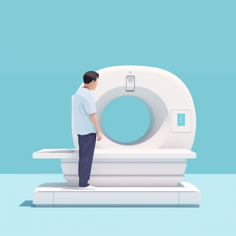
CT scans, also known as computed tomography scans, have become a critical tool in medical imaging. These scans provide detailed cross-sectional images of the body, allowing healthcare professionals to diagnose and monitor a wide range of conditions and abnormalities. The importance of CT scans in medical imaging cannot be overstated, as they provide valuable information that may not be obtainable through other imaging techniques. From identifying injuries and diseases to guiding surgical procedures, CT scans have revolutionized the practice of medicine, ultimately leading to improved patient outcomes. In this article, we will explore the various ways in which CT scans are crucial in the field of medical imaging, and why they continue to be an indispensable tool for healthcare providers.
The organization is currently facing several challenges and limitations when it comes to addressing the urgent need for digital transformation. Firstly, the technology infrastructure is outdated and not equipped to support the organization's digital transformation initiatives. Additionally, there is a lack of in-house expertise in modern digital technologies, making it difficult to implement necessary changes. Moreover, budget constraints are hindering the organization's ability to invest in new technologies and hire external expertise.
These challenges have a significant impact on the organization's ability to remain competitive in the market and meet customer demands. The outdated technology infrastructure and lack of expertise hinder the organization's ability to innovate, adapt to market changes, and deliver seamless digital experiences to customers. This, in turn, can lead to a loss of market share and decreased customer satisfaction. In order to remain competitive, it is crucial for the organization to address these challenges and invest in digital transformation.
Introduction:
Computed Tomography (CT) scans have revolutionized the field of diagnostic imaging, providing detailed images of the body's internal structures with unprecedented clarity. This technology has evolved significantly since its inception, with ongoing innovations continuing to improve its capabilities. This article will explore the history of CT scans, from its humble beginnings to its modern-day applications. It will also examine the current state of CT scans, including advancements in technology, its widespread use in medical practice, and the impact it has on patient care.
History of CT Scans:
The history of CT scans dates back to the 1970s when the first CT scanner was developed by British engineer Godfrey Hounsfield and South African physicist Allan Cormack. Their groundbreaking work earned them the Nobel Prize in Physiology or Medicine in 1979. Since then, CT technology has advanced significantly, with improvements in speed, resolution, and the ability to capture images of soft tissue, making it an invaluable tool in modern medicine.
Current State of CT Scans:
Today, CT scans are an essential diagnostic tool used in various medical specialties, including oncology, cardiology, and neurology. The availability of faster and more powerful CT scanners has led to quicker and more accurate diagnoses, facilitating better patient outcomes. Additionally, innovations such as 3D and 4D CT imaging, dual-source CT, and spectral imaging have expanded the capabilities of CT scans, making them indispensable in the management of complex medical conditions. As technology continues to evolve, the future of CT scans holds the promise of even more advanced imaging capabilities, further enhancing their role in healthcare.
Godfrey Hounsfield invented the CT scan in the early 1970s, revolutionizing the field of medical imaging. His invention allowed physicians to see inside the human body in a way that was previously impossible, providing detailed cross-sectional images of the body's internal structures.
Since its invention, CT scan technology has evolved significantly. Advancements such as faster scanning times, higher image resolution, and the ability to capture 3D images have greatly improved the diagnostic capabilities of CT scans. The development of CT angiography has also allowed for non-invasive visualization of blood vessels, aiding in the diagnosis and treatment of cardiovascular diseases.
The impact of CT scan technology on medical diagnosis and treatment has been profound. It has allowed for earlier and more accurate detection of various diseases and conditions, leading to better treatment outcomes. CT scans are now an essential tool in the diagnosis of cancer, cardiovascular diseases, and neurological disorders, among others.
Overall, the invention of the CT scan by Godfrey Hounsfield has transformed medical imaging and has played a crucial role in advancing the field of medicine. The continuous advancements in CT scan technology continue to improve the accuracy and efficiency of medical diagnosis and treatment.
Since its inception in the 1970s, CT technology has undergone significant evolution and advancements. The image quality has improved drastically over the years, with the ability to produce high-resolution, detailed images that aid in accurate diagnosis. Furthermore, advancements in CT technology have led to a reduction in radiation exposure for patients, making it a safer option for medical imaging.
In addition, the development of faster scanning capabilities has decreased the time it takes to obtain images, leading to quicker diagnoses and treatment planning. The introduction of spectral CT has allowed for more detailed and specific information to be gathered from the same scan, enhancing the diagnostic capabilities of CT imaging.
These advancements have had a profound impact on medical diagnosis and treatment planning. CT technology has become an essential tool in the detection and monitoring of various health conditions, allowing for more precise and timely interventions.
Key advantages of CT technology over traditional X-rays include its ability to provide cross-sectional images, producing detailed 3D views of the body, and its capability to distinguish between different types of soft tissues. Overall, the evolution and advancements in CT technology have revolutionized medical imaging and significantly improved patient care.
CT scans, or computed tomography scans, are widely used in healthcare for their ability to provide detailed cross-sectional images of the body. They are essential in diagnosing a variety of medical conditions, including broken bones, cancer, heart issues, and internal organ assessment. In the case of broken bones, CT scans can provide more detailed images than traditional X-rays, allowing for a more accurate diagnosis and treatment plan. In cancer diagnosis, CT scans can help detect the location and size of tumors, as well as monitor the effectiveness of treatment. Additionally, CT scans play a crucial role in assessing heart issues, such as coronary artery disease and congenital heart defects. They are also used to examine internal organs, such as the liver, lungs, and kidneys, for conditions like infections, tumors, or blood clots.
Recently, there has been a significant advancement in CT scanning technology with the integration of dark-field X-ray technology into clinical CT scanners. This technology provides additional tissue structure information, particularly in soft tissues, making it possible to visualize and characterize tissue microstructures more accurately. This advancement has the potential to improve the diagnosis and treatment of various medical conditions, further enhancing the capabilities of CT scans in healthcare.
Advancements in Image Quality and Radiation Doses have been a significant focus in the field of medical imaging. With the constant evolution of technology, healthcare providers strive to improve image resolution, clarity, and overall quality, enabling more accurate and detailed diagnoses. At the same time, reducing radiation doses during imaging procedures is a top priority to minimize potential health risks for patients. This has led to the development of innovative imaging techniques and equipment that offer high-quality images while maintaining lower radiation exposure. In this article, we will explore the latest advancements in image quality and radiation dose reduction, highlighting the benefits and impact of these developments on patient care and safety.
Image quality plays a crucial role in the accuracy of medical diagnostics. Factors such as resolution, contrast, and noise can significantly affect the interpretability of medical images. High resolution allows for sharper and more detailed images, aiding in the detection of finer abnormalities. Adequate contrast ensures that subtle differences in tissue density can be accurately identified, while minimizing noise reduces the potential for false positives or misinterpretations.
The relationship between image quality and the ability to detect subtler features and abnormalities is critical in medical imaging. Higher image quality enables healthcare professionals to identify smaller or less obvious abnormalities, leading to more accurate diagnoses and treatment plans. Inaccurate or missed diagnoses due to poor image quality can have direct implications on patient outcomes and treatment decisions.
Ultimately, the quality of medical images directly impacts the diagnostic accuracy of healthcare professionals. By providing clear and detailed images, potential abnormalities can be detected at an early stage, leading to better patient outcomes and improved overall care. Therefore, prioritizing image quality in medical diagnostics is essential for ensuring accurate and reliable interpretations.
Recent technological developments have significantly improved image resolution and clarity in CT scanning and other medical imaging systems. Advancements in CT scan technology, such as higher slice systems, have allowed for more precise and detailed imaging. These higher slice systems can capture more data in a single rotation, resulting in improved resolution and clearer images. Additionally, the introduction of handheld ultrasound scanners has allowed for more precise and accurate imaging in a variety of medical settings.
Furthermore, the history of MRI and functional MR imaging has also seen significant advancements, leading to improved image quality and clarity. These advancements have been made possible through technological developments that have enhanced speed, precision, and automation in medical imaging systems. As a result, medical professionals are now able to capture high-quality images with greater detail, leading to more accurate diagnoses and better patient care. Overall, recent technological developments in CT scanning and other medical imaging systems have greatly improved image resolution and clarity, allowing for more accurate and effective medical imaging.
Modern CT scanners have made significant advancements in reducing radiation doses to patients. One of the key developments is the use of photon counting detectors, which allow for more precise control over the amount of radiation used during imaging. These detectors can also differentiate between different energy levels of photons, leading to improved image quality with lower radiation exposure.
Additionally, filters have been incorporated into the scanners to remove softer energy radiation, further reducing the overall radiation dose. Region-of-interest imaging technology is another strategy that allows for targeted imaging of specific areas, minimizing unnecessary exposure to surrounding tissues.
Monitoring and comparing radiation doses across facilities is crucial in ensuring that proper protocols are being followed and that doses are kept as low as reasonably achievable. Appropriateness guidelines help ensure that CT scans are only performed when truly necessary, and individualized dose protocols can tailor radiation exposure to each patient's specific needs, further controlling and minimizing radiation exposure.
Overall, these advancements and strategies are essential in reducing radiation doses in modern CT scanners, ultimately prioritizing patient safety and well-being while still providing high-quality diagnostic imaging.
Three-dimensional imaging capabilities have revolutionized the way we perceive and interact with visual information across various fields such as medicine, engineering, entertainment, and art. With the ability to capture and display data in three dimensions, this technology allows for more immersive and accurate visualization of objects and spaces. From medical imaging techniques like CT and MRI scans to virtual reality experiences and 3D modeling for architecture and design, three-dimensional imaging has opened up a whole new world of possibilities. In this article, we will explore the different applications and advancements in three-dimensional imaging capabilities, and how they are shaping the future of various industries.
Three-dimensional (3D) images play a crucial role in medical diagnosis and treatment planning by providing detailed and accurate information about internal structures. These images allow medical professionals to visualize the anatomy in a more comprehensive way, leading to more precise assessments and diagnoses. They are particularly valuable in identifying and understanding complex conditions such as tumors, fractures, and malformations.
When it comes to treatment planning, 3D images are essential for mapping out surgical procedures with high precision. By accurately visualizing the area of interest, surgeons can better plan their approach and anticipate potential challenges, ultimately reducing the risk of complications during the operation. Additionally, these images can aid in determining the most effective course of treatment, such as choosing the appropriate size and placement of implants or designing personalized medical devices.
Overall, the use of 3D images in medical diagnosis and treatment planning improves the accuracy of assessments and interventions. This not only enhances patient outcomes but also reduces the likelihood of errors and complications during surgical procedures. As technology continues to advance, the integration of 3D imaging in medical practice will continue to play a critical role in improving patient care.
Techniques used to generate three-dimensional images from CT scans include the integration of dark-field X-ray method, conventional X-ray technology, and photon counting CT technology. The dark-field X-ray method allows for the visualization of small structures, such as lung tissue, that are not easily detectable with conventional X-ray technology. The integration of both methods provides more detailed and accurate three-dimensional images.
Additionally, photon counting CT technology is used to count and measure individual photons, resulting in improved image quality and lower radiation dose. This technique allows for better visualization of soft tissue and small structures, and it has the potential to enhance diagnostic capabilities.
Furthermore, the process of combining multiple X-ray images is utilized to create three-dimensional images. By capturing multiple X-ray images from different angles, a computer algorithm reconstructs them into a three-dimensional representation, providing a comprehensive view of the scanned area.
In conclusion, the integration of dark-field X-ray, conventional X-ray technology, and photon counting CT technology, as well as the combination of multiple X-ray images, are essential techniques used to generate high-quality three-dimensional images from CT scans.
Computed Tomography (CT) technology has seen significant advancements in recent years, leading to improved diagnosis and treatment for patients. From faster scanning times to enhanced image quality, recent innovations in CT technology are providing healthcare professionals with the tools they need to deliver superior care to their patients. In this article, we will explore some of the most notable advancements in CT technology and their impact on the medical field. These innovations are revolutionizing the way medical professionals diagnose and treat a wide range of conditions, ultimately improving patient outcomes and advancing the field of healthcare.
Photon-counting detectors are a revolutionary technology in CT imaging, enhancing image acquisition by counting and measuring individual X-ray photons. Unlike traditional energy-integrating detectors, photon-counting detectors provide detailed spectral information, allowing for better spatial resolution and image contrast. This technology enables the differentiation of different energy levels of photons, resulting in more accurate and precise images.
Additionally, photon-counting detectors have the potential to reduce electronic noise and radiation dose, aligning with the goals of Image Wisely. By accurately measuring each photon's energy, these detectors can effectively filter out electronic noise, leading to clearer images with improved diagnostic accuracy. Furthermore, the ability to distinguish between high and low-energy photons can potentially reduce radiation dose, making CT imaging safer for patients.
In conclusion, photon-counting detectors have the potential to significantly improve image acquisition in CT imaging, offering detailed spectral information for better spatial resolution and image contrast, while also supporting the principles of Image Wisely by reducing electronic noise and radiation dose.